|
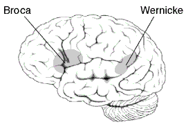
9/30/2018 0 Comments I Don't quite comprehend...The idea of localization that was introduced by Paul Broca continued to be debated and analyzed by neuroscientists. New ideas that left and right hemispheres of the brain were more dominant in certain functions became more accepted with additional support from Carl Wernicke’s work. Wernicke's areaLOCATION The Wernicke’s area is located on the left hemisphere of the brain. Just like the Broca’s area, it is located on the left hemisphere of the brain, which plays a major role in the production and interpretation of language. This area encompasses the Sylvian fissure, which is a diagonal chasm that borders the parietal and temporal lobes. THE STORY In 1874, just over a decade after Broca, Carl Wernicke also shared Broca’s idea of localization. This belief was furthered by one of Wernicke's patients who had endured a stroke. His patient was able to engage in speech and showed no sign of auditory impairments. However, his language comprehension was lacking. He was unable to recognize written or verbal speech. After the death of his patient, Wernicke explored the deceased’s brain, finding a lesion at the junction on the posterior of the temporal and parietal lobes. From this discovery, Wernicke linked the lesion to the man’s poor comprehension, calling the impairment “sensory aphasia.” FUNCTION The Wernicke’s area is in charge of language comprehension. It plays a major role in semantics, form, and pragmatics. This is the sensory area of the brain, since it receives and interprets external information. WERNICKE’S APHASIA Today, “sensory aphasia” is now known as Wernicke’s aphasia or sometimes referred to as fluent aphasia. In Wernicke’s discovery, this aphasia appeared to be articulate language, but the structure and meaning behind words made little sense. In a video example, which can be observed below, a client with Wernicke’s aphasia is asked by a clinician, “What is this?” as she gestures to her mouth. The patient responds with, "the end of a football,” and continues to back up his answer with other properly articulated words. Unlike Broca’s aphasia, the trouble comes from the lack of meaning of what the afflicted individual is expressing. TAkeaway 🎈The Wernicke’s area is also on the left hemisphere of the brain like the Broca’s area, since this hemisphere is language dominant. Unlike Broca’s aphasia, Wernicke’s aphasia deals with an individual’s cognitive processing. Next week
0 Comments
9/21/2018 0 Comments It's at the tip of my tongue!The brain is a complex organ that relies on communication between all of its different areas, making it difficult to localize where certain functions stem from. However, this idea of localization started with Paul Broca’s discovery that harming a particular area of the brain resulted in a speech impairment. Broca's AreaLOCATION The Broca’s area is located in the frontal lobe or more specifically in the region known as the inferior gyrus. This area is also thought to reside in the left hemisphere of the brain. As previously discussed, the left hemisphere plays a major role in analytical reasoning, but language is also dominant on this side. THE STORY In 1861, Paul Pierre Broca received a patient who had a major infection in his leg. The wound had already begun to slightly decompose, and Broca had little faith in the man’s recovery, let alone survival. However, this man also had a peculiar disorder that was unknown at its time, but would later be described as aphasia, or in this case Broca’s aphasia. Broca’s patient, Louis Victor Leborgne, struggled with the motor production of language. Broca could see the frustration that Leborgne faced, while trying to use his limited speech to convey messages. It was noted that the one of the only consistent words Leborgne could produce was “tan.” There was a clear desire to express his thoughts, but the production was not eminent. Later on, Leborgne died and Broca saw this as an opportunity to see if localization theories of the brain could be true. He did an examination of the brain and discovered a cavity the size of “a chicken’s egg” in Leborgne’s frontal lobe. Interestingly enough, this damage was located on the patient’s left side of the brain. Broca continued to look for more clients like Leborgne and found that each time, there was some kind of damage to the left frontal lobe of the brain. This area became known as the Broca’s area. FUNCTION Broca’s area is most tied to expressive language. It is linked to “language comprehension” and motor movements, as well as “sensorimotor learning and integration.” BROCA’S APHASIA Louis Victor Leborgne was diagnosed with Broca’s aphasia, since he spoke using telegraphic speech and low level syntax structure. He was unable to build full-fledged sentences, and even if he knew what he wanted to say, Leborgne was unable to verbalize his idea using standard rules of language. The best way I can think to describe Broca’s aphasia is when you can’t remember a word, but you are able to describe what the word is. Even though you know the idea of the word, you still cannot recall the specific word that you are visualizing in your mind. This experience is when a word is at the “tip of your tongue." You have an idea of what you want to say, yet you are still unable to vocalize that to your peers. Now think of that on a much larger scale. People with Broca’s aphasia have thoughts and ideas, but it’s their motor planning that inhibits their thoughts to become speech. Takeaway 🎈The Broca's area is located on the left frontal lobe of the brain, and its prime function is to allow expressive language. When this area becomes damaged it can result in Broca’s aphasia, which causes an individual to use telegraphic speech. Next week We will dive into another speech localized area of the brain and learn about a different kind of aphasia.
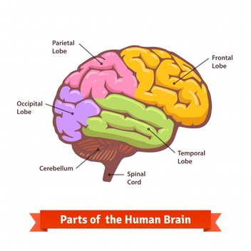
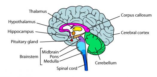
Links: psychology.jrank.org/pages/384/Localization-Brain-Function.html www.neuroscientificallychallenged.com/blog/know-your-brain-brocas-area www.britannica.com/science/Broca-area Images: http://www.cerebromente.org.br/n02/historia/areabroca.gif 9/14/2018 0 Comments A Brain overviewEvery Speech-Language Pathologist, or future SLP should have some knowledge of human anatomy. Yes, the eyes, ears, and mouth are important parts when hearing and understanding speech. However, these parts receive and execute functions that stem from a vital organ. The brain. The brain is the command center for everything in your body, and without it you would not be alive, let alone able to talk. Before I can focus on speech-related areas of the brain and what happens when brain development does not go according to plan, it is important to have a brief review of the basic anatomy of the brain. Definitions
This was just a brief overview of the parts of the brain. If you want to read more on each individual part, I will paste the links I referenced down below. Takeaway 🎈The most important concept to take away is that the brain is made up of all these very specific parts, but together they work together and and form neural connections that allow us to execute basic and detailed tasks. The brain is the first organ that begins to develop in a fetus and is necessary for other systems of the body to work, since it oversees a person’s physicality and mentality.
Links: https://mayfieldclinic.com/pe-anatbrain.htm https://www.livescience.com/29365-human-brain.html Images: https://www.freepik.com/free-vector/doodle-birthday-elements_903451.htm https://www.freepik.com/free-photos-vectors/flower https://www.freepik.com/free-photos-vectors/background https://image.freepik.com/free-vector/colored-and-labeled-human-brain-diagram_3446-148.jpg https://media.proprofs.com/images/QM/user_images/1826446/1494570168.jpg |
 RSS Feed
